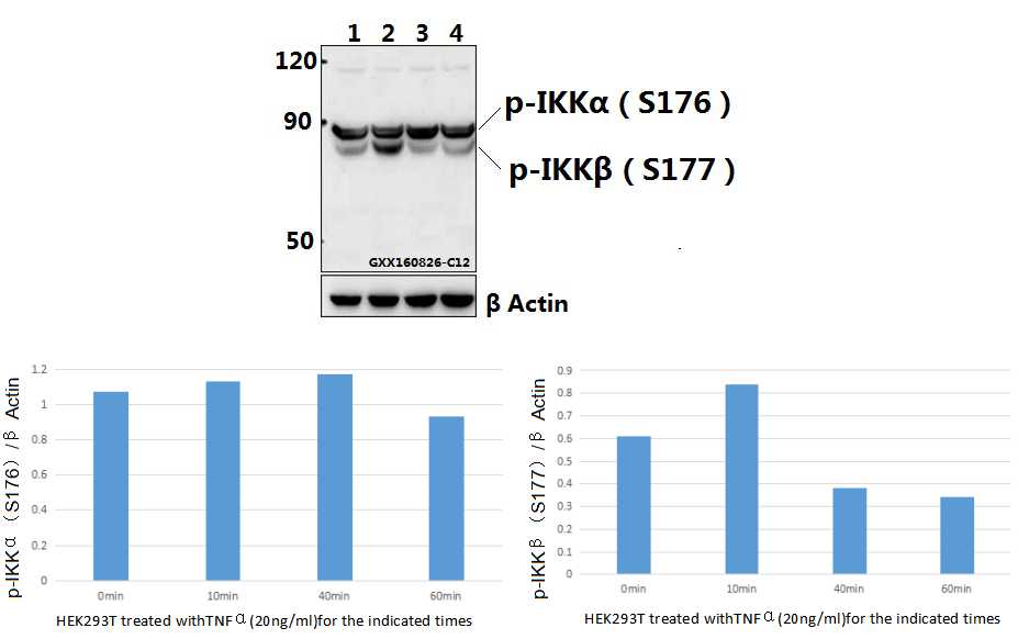
Western blot (WB) analysis of IKKα/β (phospho-S176/177) polyclonal antibody at 1:500 dilution
Lane1:HEK293T whole cell lysate
Lane2:HEK293T treated with TNFα(20ng/ml)for 10 minutes whole cell lysate
Lane3:HEK293T treated with TNFα(20ng/ml)for 40 minutes whole cell lysate
Lane4:HEK293T treated with TNFα(20ng/ml)for 1 hour whole cell lysate
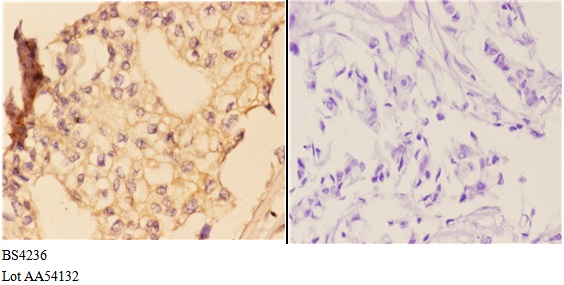
Immunohistochemistry (IHC) analyzes of p-IKKα/β (S176/177) pAb in paraffin-embedded human breast carcinoma tissue at 1:50,showing cytoplasmic and nuclear staining.Negative control (the right)Using PBS instead of primary antibody, secondary antibody is Goat Anti-Rabbit IgG-biotin followed by avidin-peroxidase.
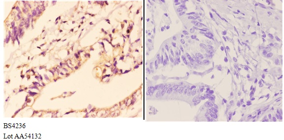
Immunohistochemistry (IHC) analyzes of p-IKKα/β (S176/177) pAb in paraffin-embedded human colon carcinoma tissue at 1:50,showing cytoplasmic and nuclear staining.Negative control (the right)Using PBS instead of primary antibody, secondary antibody is Goat Anti-Rabbit IgG-biotin followed by avidin-peroxidase.
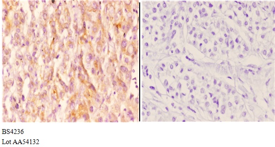
Immunohistochemistry (IHC) analyzes of p-IKKα/β (S176/177) pAb in paraffin-embedded human liver carcinoma tissue at 1:50,showing cytoplasmic and nuclear staining.Negative control (the right)Using PBS instead of primary antibody, secondary antibody is Goat Anti-Rabbit IgG-biotin followed by avidin-peroxidase.