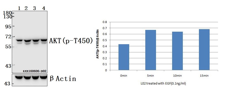
Western blot (WB) analysis of AKT (phospho-T450) polyclonal antibody at 1:500 dilution
Lane1:L02 whole cell lysate
Lane2:L02 treated with EGF(0.1ng/ml,5min) whole cell lysate
Lane3:L02 treated with EGF(0.1ng/ml,10min) whole cell lysate
Lane4:L02 treated with EGF(0.1ng/ml,15min) whole cell lysate
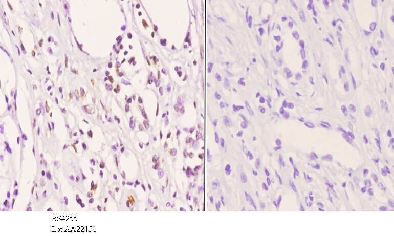
Immunohistochemistry (IHC) analyzes of p-AKT (T450) pAb in paraffin-embedded human kidney carcinoma tissue at 1:50.showing cytoplasmic and nucleus staining. Negative control (the right)Using PBS instead of primary antibody, secondary antibody is Goat Anti-Rabbit IgG-biotin followed by avidin-peroxidase.
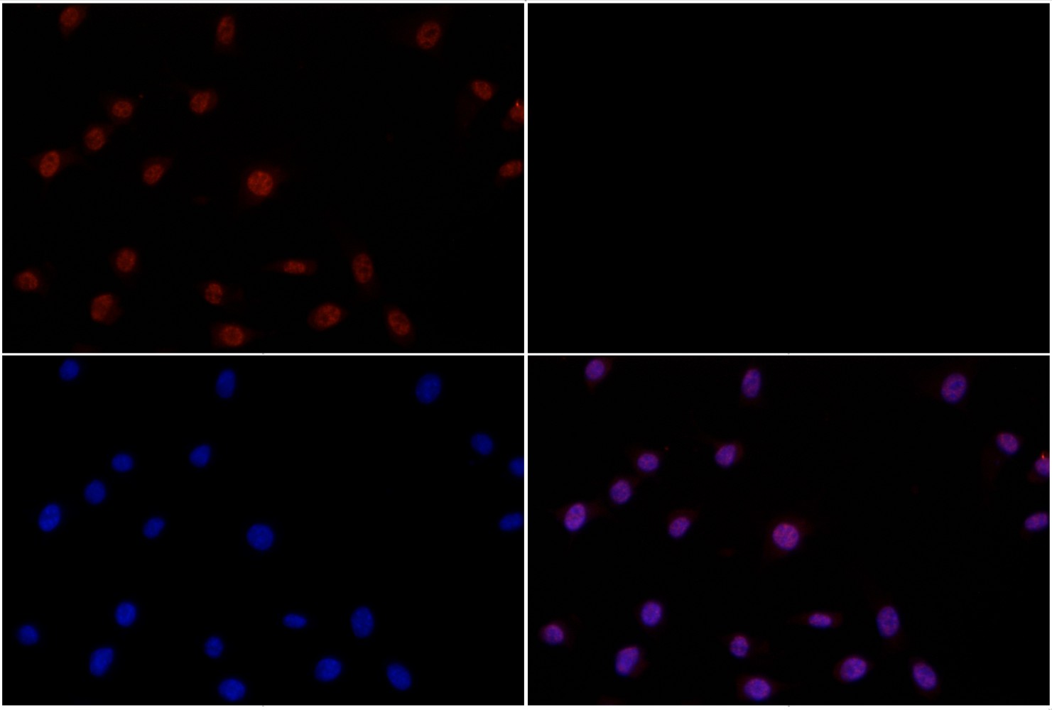
Immunofluorescence analysis of NIH-3T3 cells using AKT (phospho-T450) pAb at dilution of 1:200 (40x lens).
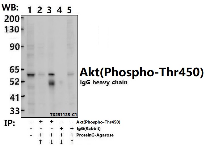
Immunoprecipitation of 3T3-L1 cell lysates using AKT (phospho-T450) pAb (Sepharose Bead Conjugate)#BD0048 (lane 2 and lane 3) and Nonspecific IgG Control (Sepharose Bead Conjugate)#BD0048 (lane 4 and lane 5) .Lane 1 is 30% input. The western blot was probed using AKT (phospho-T450) pAb.