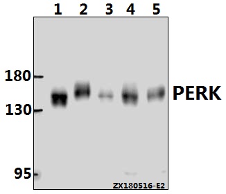
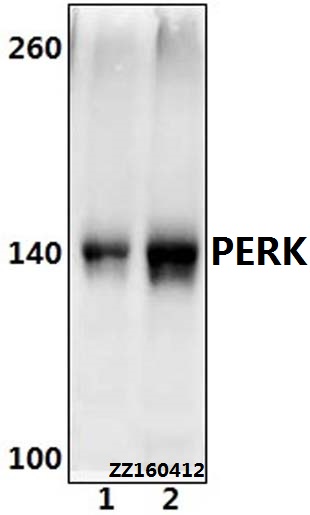
Western blot (WB) analysis of PERK (R87) pAb at 1:1000 dilution
Lane1:HCT116 whole cell lysate(20ug)
Lane2:MCF-7 whole cell lysate(20ug)
Lane3:A375 whole cell lysate(20ug)
Lane4:CT26 whole cell lysate(40ug)
Lane5:PMVEC whole cell lysate(40ug)
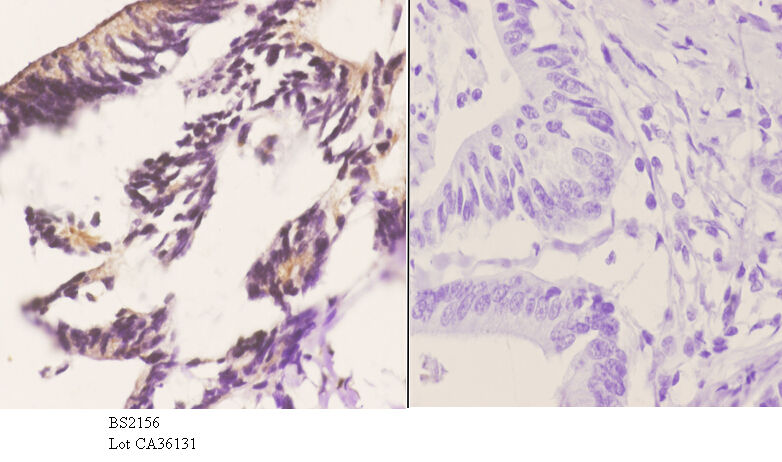
Immunohistochemistry (IHC) analyzes of PERK (R87) pAb in paraffin-embedded human colon carcinoma tissue at 1:50.showing cytoplasmic and nucleus staining. Negative control (the right)Using PBS instead of primary antibody, secondary antibody is Goat Anti-Rabbit IgG-biotin followed by avidin-peroxidase.
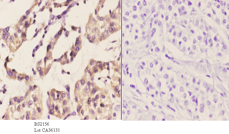
Immunohistochemistry (IHC) analyzes of PERK (R87) pAb in paraffin-embedded human liver carcinoma tissue at 1:50.showing cytoplasmic and nucleus staining. Negative control (the right)Using PBS instead of primary antibody, secondary antibody is Goat Anti-Rabbit IgG-biotin followed by avidin-peroxidase.