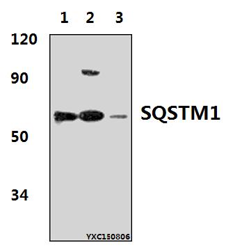
Western blot (WB) analysis of SQSTM1 polyclonal antibody at 1:500 dillution
Lane1:Hela whole cell lysate(40μg)
Lane2:NIH-3T3 whole cell lysate(30μg)
Lane3:PC12 whole cell lysate(40μg)
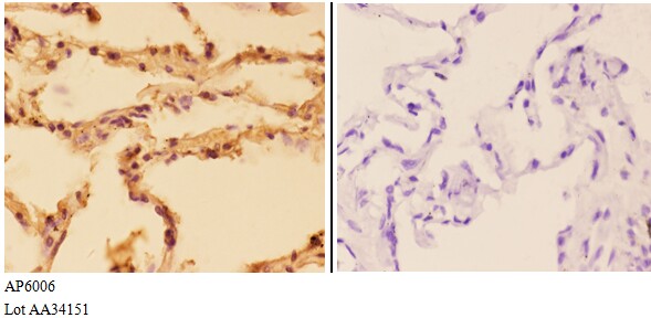
Immunohistochemistry (IHC) analyzes of SQSTM1 pAb in paraffin-embedded human lung carcinoma tissue at 1:50,showing cytoplasm staining.Negative control (the right)Using PBS instead of primary antibody, secondary antibody is Goat Anti-Rabbit IgG-biotin followed by avidin-peroxidase.
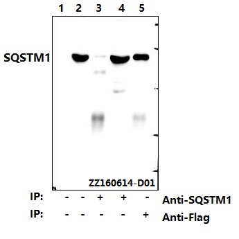
Western blot (WB) analysis of SQSTM1 pAb at 1:500 dilution
Lane1:HEK293T whole cell lysate ,untransfected
Lane2:HEK293T whole cell lysate , transfected with pcDNA3.1-HA-FlaG-p62(human)
Lane3:HEK293T whole cell lysate , transfected with pcDNA3.1-HA-FlaG-p62(Anti-SQSTM1, IP)
Lane4:HEK293T whole cell lysate , transfected with pcDNA3.1-HA-FlaG-p62(Anti-SQSTM1, IP,supernatant)
Lane5:HEK293T whole cell lysate , transfected with pcDNA3.1-HA-FlaG-p62(Anti-Flag, IP)
Immunofluorescence analysis of U-8MG cells using SQSTM1 antibody at dilution of 1:50.