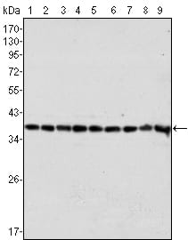
Western blot analysis using GAPDH mouse mAb against Hela (1), A549 (2), A431 (3), MCF-7 (4), K562 (5), Jurkat (6), HL60 (7), SKN-SH (8) and SKBR-3 (9) cell lysate.
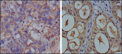
Immunohistochemical analysis of paraffin-embedded human breast carcinoma (left) and kidney carcinoma (right), showing cytoplasmic localization using GAPDH mouse mAb with DAB staining.
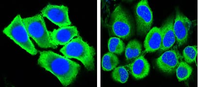
Confocal Immunofluorescence analysis of methanol-fixed HepG2 (left) and Hela (right) cells using GAPDH mouse mAb (green), showing cytoplasmic localization. Blue: DRAQ5 fluorescent DNA dye.
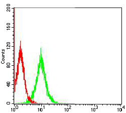
Flow cytometric analysis of NIH3T3 cells using GAPDH mouse mAb (green) and negative control (red).