中国 EN 400-660-9091

联系我们

电话:025-68037686

地址:江苏生命科技创新园F6幢1层

订购:nanjing03@biogot.com

服务:biorase01@biogot.com

合作:lvyun@biogot.com

支持:may@biogot.com

ZAP70 monoclonal antibody

货号: MB66987
价格 1350
货期 现货(1-2个工作日)
25ul 50ul 100ul 200ul
No. :MB66987
Product Name :ZAP70 monoclonal antibody
Swiss-Prot :P43403
Host :Mouse
Reactivity :Human, Mouse
Applications :WB, IF/ICC, FC
Application_all :WB (1/500 - 1/1000), IF/ICC (1/10 - 1/50), FC (1/10 - 1/50)
Background :The Syk family protein tyrosine kinase Zap-70 is expressed in T and NK cells and plays a critical role in mediating T cell activation in response to T cell receptor (TCR) engagement . Following TCR engagement, Zap-70 is rapidly phosphorylated on several tyrosine residues through autophosphorylation and transphosphorylation by the Src family tyrosine kinase Lck . Tyrosine phosphorylation correlates with increased Zap-70 kinase activity and downstream signaling events. Expression of Zap-70 is correlated with disease progression and survival in patients with chronic lymphocytic leukemia .
Product :Mouse IgG2a kappa. Liquid in PBS, pH 7.3, 30% glycerol, and 0.01% sodium azide.
Purification&Purity :This antibody is purified through a protein G column.
Storage&Stability :Store at 4°C short term. Aliquot and store at -20°C long term. Avoid freeze-thaw cycles.
Specificity :Recognizes endogenous levels of ZAP70 protein.
BiowMW :~ 70 kDa
Note :For research use only, not for use in diagnostic procedure.
Pathway :T cell receptor signaling.
Alternative Name :SRK; Tyrosine-protein kinase ZAP-70; 70 kDa zeta-chain associated protein; Syk-related tyrosine kinase
Immunogen :Recombinant fusion protein of human ZAP70. The exact sequence is proprietary.
Conjugate :Unconjugated
Modification :Unmodification
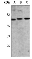
Western blot analysis of ZAP70 expression in Jurkat (A), MOLT4 (B), mouse thymus (C) whole cell lysates.
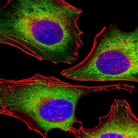
Immunofluorescent analysis of ZAP70 staining in Hela cells. Formalin-fixed cells were permeabilized with 0.1% Triton X-100 in TBS for 5-10 minutes and blocked with 3% BSA-PBS for 30 minutes at room temperature. Cells were probed with the primary antibody in 3% BSA-PBS and incubated overnight at 4 °C in a humidified chamber. Cells were washed with PBST and incubated with a AF488-conjugated secondary antibody (green) in PBS at room temperature in the dark. Phalloidin - AF555 was used to stain the cytoplasm (red). DAPI was used to stain the cell nuclei (blue).

关注我们

400-660-9091

电话:025-68037686

地址:江苏生命科技创新园F6幢1层

订购:nanjing03@biogot.com

服务:biorase01@biogot.com

合作:lvyun@biogot.com

支持:may@biogot.com