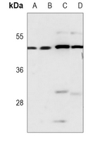
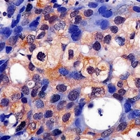
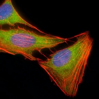
Product Name :ERK1 monoclonal antibody
Applications :WB, IHC, IF/ICC
Application_all :WB (1/500 - 1/1000), IHC (1/50 - 1/200), IF/ICC (1/10 - 1/50)
Background :Mitogen-activated protein kinases (MAPKs) are a widely conserved family of serine/threonine protein kinases involved in many cellular programs, such as cell proliferation, differentiation, motility, and death. The p44/42 MAPK (Erk1/2) signaling pathway can be activated in response to a diverse range of extracellular stimuli, including mitogens, growth factors, and cytokines , and research investigators consider it an important target in the diagnosis and treatment of cancer . Upon stimulation, a sequential three-part protein kinase cascade is initiated, consisting of a MAP kinase kinase kinase (MAPKKK or MAP3K), a MAP kinase kinase (MAPKK or MAP2K), and a MAP kinase (MAPK). Multiple p44/42 MAP3Ks have been identified, including members of the Raf family, as well as Mos and Tpl2/COT. MEK1 and MEK2 are the primary MAPKKs in this pathway . MEK1 and MEK2 activate p44 and p42 through phosphorylation of activation loop residues Thr202/Tyr204 and Thr185/Tyr187, respectively. Several downstream targets of p44/42 have been identified, including p90RSK and the transcription factor Elk-1 . p44/42 are negatively regulated by a family of dual-specificity (Thr/Tyr) MAPK phosphatases, known as DUSPs or MKPs , along with MEK inhibitors, such as U0126 and PD98059.
Product :Mouse IgG1 kappa. Liquid in PBS, pH 7.3, 30% glycerol, and 0.01% sodium azide.
Purification&Purity :This antibody is purified through a protein G column.
Storage&Stability :Store at 4°C short term. Aliquot and store at -20°C long term. Avoid freeze-thaw cycles.
Specificity :Recognizes endogenous levels of ERK1 protein.
Note :For research use only, not for use in diagnostic procedure.
Alternative Name :ERK1; PRKM3; Mitogen-activated protein kinase 3; MAP kinase 3; MAPK 3; ERT2; Extracellular signal-regulated kinase 1; ERK-1; Insulin-stimulated MAP2 kinase; MAP kinase isoform p44; p44-MAPK; Microtubule-associated protein 2 kinase; p44-ERK1
Immunogen :KLH-conjugated synthetic peptide encompassing a sequence within the center region of human ERK1. The exact sequence is proprietary.
Modification :Unmodification