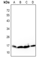
Western blot analysis of Histone H3 expression in Hela (A), Raw264.7 (B), mouse brain (C), rat brain (D) whole cell lysates.
Immunofluorescence analysis of NIH/3T3 cells using AKR1C3 antibody at dilution of 1:100. Blue: DAPI for nuclear staining.
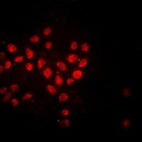
Immunofluorescent analysis of Histone H3 staining in Hela cells. Formalin-fixed cells were permeabilized with 0.1% Triton X-100 in TBS for 5-10 minutes and blocked with 3% BSA-PBS for 30 minutes at room temperature. Cells were probed with the primary antibody in 3% BSA-PBS and incubated overnight at 4 °C in a hidified chamber. Cells were washed with PBST and incubated with a DyLight 594-conjugated secondary antibody (red) in PBS at room temperature in the dark.