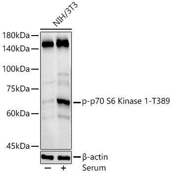
Western blot analysis of NIH/3T3, using Phospho-p70 S6 Kinase 1-T389 antibody at 1:500 dilution.NIH/3T3 cells were treated by 10% FBS at 37℃ for 30 minutes after serum-starvation overnight.
Secondary antibody: HRP Goat Anti-Rabbit IgG at 1:10000 dilution.
Lysates/proteins: 25ug per lane.
Blocking buffer: 3% nonfat dry milk in TBST.
Detection: ECL Basic Kit .
Exposure time: 180s.
电话:025-68037686
地址:江苏生命科技创新园F6幢1层
订购:nanjing03@biogot.com
服务:biorase01@biogot.com
合作:lvyun@biogot.com
支持:may@biogot.com
