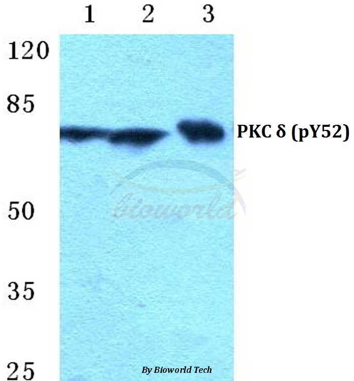
Western blot (WB) analysis of p-PKC δ (Y52) polyclonal antibody at 1:500 dilution
Lane1:MCF-7 cell lysate treated with serum starvation(24h)
Lane2:Raw264.7 cell lysate treated with serum starvation(24h)
Lane3:H9C2 cell lysate treated with serum starvation(24h)
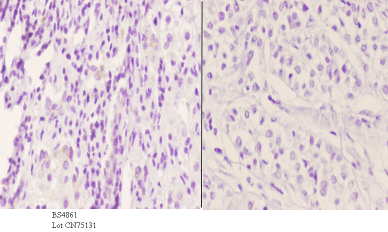
Immunohistochemistry (IHC) analyzes of p-PKC δ (Y52) pAb in paraffin-embedded human liver carcinoma tissue at 1:50.showing cytoplasmic and nucleus staining. Negative control (the right)Using PBS instead of primary antibody, secondary antibody is Goat Anti-Rabbit IgG-biotin followed by avidin-peroxidase.