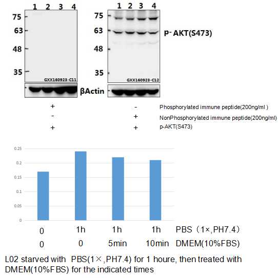
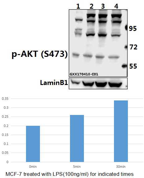
Western blot (WB) analysis of AKT (phospho-S473) polyclonal antibody at 1:500 dilution
Lane1:L02 whole cell lysate
Lane2:L02 starved with PBS(1×PBS,PH7.4) for 1 houre whole cell lysate
Lane3:L02 starved with PBS(1×PBS,PH7.4) for 1 houre then treated with DMEM(10%FBS) for 5 minutes whole cell lysate
Lane4:L02 starved with PBS(1×PBS,PH7.4) for 1 houre then treated with DMEM(10%FBS) for 10 minutes whole cell lysate
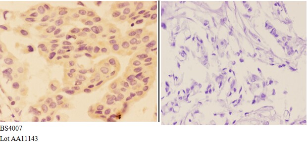
Immunohistochemistry (IHC) analyzes of AKT (phospho-S473) pAb in paraffin-embedded human breast carcinoma tissue at 1:50,showing cytoplasm and nucleus staining.Negative control (the right)Using PBS instead of primary antibody, secondary antibody is Goat Anti-Rabbit IgG-biotin followed by avidin-peroxidase.
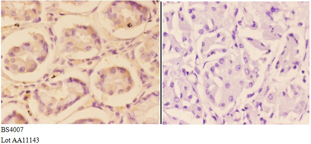
Immunohistochemistry (IHC) analyzes of AKT (phospho-S473) pAb in paraffin-embedded human stomach carcinoma tissue at 1:50,showing cytoplasm and nucleus staining.Negative control (the right)Using PBS instead of primary antibody, secondary antibody is Goat Anti-Rabbit IgG-biotin followed by avidin-peroxidase.