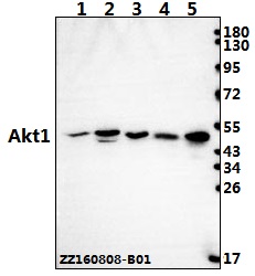
Western blot (WB) analysis of AKT1 (P125) polyclonal antibody at 1:500 dilution
Lane1:A549 whole cell lysate(40ug)
Lane2:NIH-3T3 whole cell lysate(40ug)
Lane3:PC12 whole cell lysate(40ug)
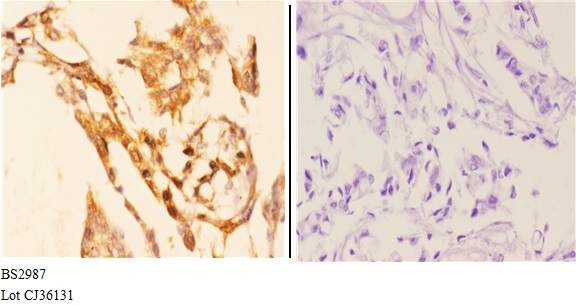
Immunohistochemistry (IHC) analyzes of AKT1 (P125) pAb in paraffin-embedded human breast carcinoma tissue at 1:50.showing Cytoplasm, Nucleus and Cell membrane staining. Negative control (the right)Using PBS instead of primary antibody, secondary antibody is Goat Anti-Rabbit IgG-biotin followed by avidin-peroxidase.
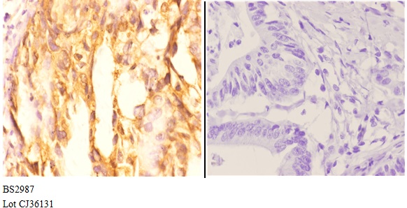
Immunohistochemistry (IHC) analyzes of AKT1 (P125) pAb in paraffin-embedded human colon carcinoma tissue at 1:50.showing Cytoplasm, Nucleus and Cell membrane staining. Negative control (the right)Using PBS instead of primary antibody, secondary antibody is Goat Anti-Rabbit IgG-biotin followed by avidin-peroxidase.
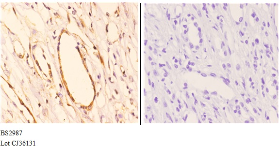
Immunohistochemistry (IHC) analyzes of AKT1 (P125) pAb in paraffin-embedded human kidney carcinoma tissue at 1:50.showing Cytoplasm, Nucleus and Cell membrane staining. Negative control (the right)Using PBS instead of primary antibody, secondary antibody is Goat Anti-Rabbit IgG-biotin followed by avidin-peroxidase.