Western blot (WB) analysis of PKAα/β cat (K17) polyclonal antibody at 1:500 dilution
Lane1:The brain tissue lysate of Rat(40ug)
Lane2:The Testis tissue lysate of Mouse(40ug)
Lane3:The Testis tissue lysate of Rat(40ug)
Lane4:MCF-7 whole cell lysate(40ug)
Lane5:HEK293T whole cell lysate(40ug)
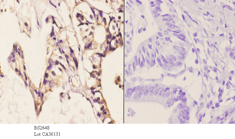
Immunohistochemistry (IHC) analyzes of PKAα/β cat (K17) pAb in paraffin-embedded human colon carcinoma tissue at 1:50.showing cytoplasmic and nucleus staining. Negative control (the right)Using PBS instead of primary antibody, secondary antibody is Goat Anti-Rabbit IgG-biotin followed by avidin-peroxidase.
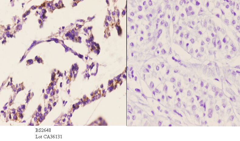
Immunohistochemistry (IHC) analyzes of PKAα/β cat (K17) pAb in paraffin-embedded human liver carcinoma tissue at 1:50.showing cytoplasmic and nucleus staining. Negative control (the right)Using PBS instead of primary antibody, secondary antibody is Goat Anti-Rabbit IgG-biotin followed by avidin-peroxidase.
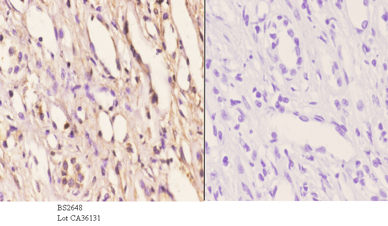
Immunohistochemistry (IHC) analyzes of PKAα/β cat (K17) pAb in paraffin-embedded human kidney carcinoma tissue at 1:50.showing cytoplasmic and nucleus staining. Negative control (the right)Using PBS instead of primary antibody, secondary antibody is Goat Anti-Rabbit IgG-biotin followed by avidin-peroxidase.