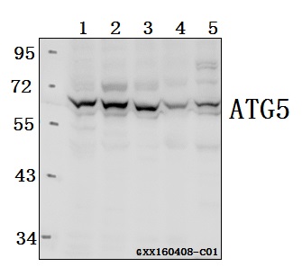
Western blot (WB) analysis of ATG 5 polyclonal antibody at 1:1000 dilution
Lane1:HCT116 whole cell lysate(40ug)
Lane2:MCF-7 whole cell lysate(30ug)
Lane3:SK-OVCAR3 whole cell lysate(40ug)
Lane4:C6 whole cell lysate(40ug)
Lane5:The brain tissue lysate of Mouse(40ug)
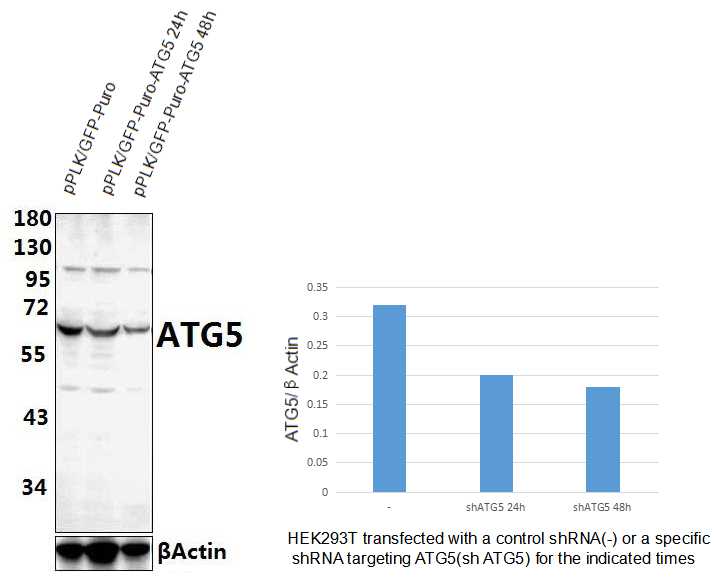
Western blot analysis of extracts from HEK293T cells transfected with control shRNA (-) (Lane 1) or ATG5 shRNA (+)(Lane 2-3). ATG5 was detected using ATG5 pAb #AP6026.The ATG5 Antibody confirms silencing of ATG5 expression.
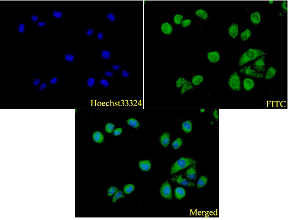
IF image of AP6026 stained A549 cells.The cells were 4% paraformaldehyde fixed (20 min) and then incubated in 10% normal goat serum for 1h to permeabilise the cells and block non-specific protein-protein interactions. The cells were then incubated with the antibody ATG5 #AP6026(1:200) at 5µg/ml overnight at +4°C. The secondary antibody (Green) was Goat Anti-Rabbit IgG (H+L) FITC #BS10950 used at a 1/1000 dilution for 1h. Hoechst33342 #BD5011 was used to stain the cell nuclei (blue).