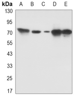
Product Name :PKC beta Rabbit monoclonal antibody
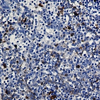
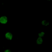
Applications :WB, IHC, IF/ICC, IP
Application_all :WB (1/500 - 1/1000), IHC (1/50 - 1/100), IF/ICC (1/50 - 1/100), IP (1/10 - 1/50)
Background :Activation of protein kinase C (PKC) is one of the earliest events in a cascade that controls a variety of cellular responses, including secretion, gene expression, proliferation, and muscle contraction . PKC isoforms belong to three groups based on calcium dependency and activators. Classical PKCs are calcium-dependent via their C2 domains and are activated by phosphatidylserine (PS), diacylglycerol (DAG), and phorbol esters (TPA, PMA) through their cysteine-rich C1 domains. Both novel and atypical PKCs are calcium-independent, but only novel PKCs are activated by PS, DAG, and phorbol esters . Members of these three PKC groups contain a pseudo-substrate or autoinhibitory domain that binds to substrate-binding sites in the catalytic domain to prevent activation in the absence of cofactors or activators. Control of PKC activity is regulated through three distinct phosphorylation events. Phosphorylation occurs in vivo at Thr500 in the activation loop, at Thr641 through autophosphorylation, and at the carboxy-terminal hydrophobic site Ser660. Atypical PKC isoforms lack hydrophobic region phosphorylation, which correlates with the presence of glutamic acid rather than the serine or threonine residues found in more typical PKC isoforms. The enzyme PDK1 or a close relative is responsible for PKC activation. A recent addition to the PKC superfamily is PKCμ (PKD), which is regulated by DAG and TPA through its C1 domain. PKD is distinguished by the presence of a PH domain and by its unique substrate recognition and Golgi localization. PKC-related kinases (PRK) lack the C1 domain and do not respond to DAG or phorbol esters. Phosphatidylinositol lipids activate PRKs, and small Rho-family GTPases bind to the homology region 1 (HR1) to regulate PRK kinase activity.PKCβ has two isoforms, PKCβI and PKCβII, due to alternative splicing.
Product :Liquid in 50mM Tris-Glycine (pH 7.4), 0.15M NaCl, 50% Glycerol, 0.01% Sodium azide and 0.05% BSA.
Purification&Purity :The antibody was purified by immunogen affinity chromatography.
Storage&Stability :Store at 4°C short term. Aliquot and store at -20°C long term. Avoid freeze-thaw cycles.
Specificity :Recognizes endogenous levels of PKC beta protein.
Note :For research use only, not for use in diagnostic procedure.
Pathway :Regulation of Apoptosis,ErbB HER Signaling,G Protein-coupled Receptors Signaling to MAPK Erk,MAPK Erk in Growth and Differentiation Pathway,Inhibition of Apoptosis,Mitochondrial Control of Apoptosis,Phospholipase Signaling,Protein Kinase C Signaling
Alternative Name :PKCB; PRKCB1; Protein kinase C beta type; PKC-B; PKC-beta
Immunogen :A synthesized peptide derived from human PKC beta 2
Modification :Unmodification