中国 EN 400-660-9091

联系我们

电话:025-68037686

地址:江苏生命科技创新园F6幢1层

订购:nanjing03@biogot.com

服务:biorase01@biogot.com

合作:lvyun@biogot.com

支持:may@biogot.com

p53 (Phospho-S6) Rabbit monoclonal antibody

货号: MB66419
价格 1350
货期 现货(1-2个工作日)
25ul 50ul 100ul 200ul
No. :MB66419
Product Name :p53 (Phospho-S6) Rabbit monoclonal antibody
Swiss-Prot :P04637
Host :Rabbit
Reactivity :Human
Applications :WB, IHC, IP
Application_all :WB (1/500 - 1/1000), IHC (1/50 - 1/100), IP (1/10 - 1/50)
Background :The p53 tumor suppressor protein plays a major role in cellular response to DNA damage and other genomic aberrations. Activation of p53 can lead to either cell cycle arrest and DNA repair or apoptosis. p53 is phosphorylated at multiple sites in vivo and by several different protein kinases in vitro. DNA damage induces phosphorylation of p53 at Ser15 and Ser20 and leads to a reduced interaction between p53 and its negative regulator, the oncoprotein MDM2 . MDM2 inhibits p53 accumulation by targeting it for ubiquitination and proteasomal degradation. p53 can be phosphorylated by ATM, ATR, and DNA-PK at Ser15 and Ser37. Phosphorylation impairs the ability of MDM2 to bind p53, promoting both the accumulation and activation of p53 in response to DNA damage. Chk2 and Chk1 can phosphorylate p53 at Ser20, enhancing its tetramerization, stability, and activity . p53 is phosphorylated at Ser392 in vivo and by CAK in vitro. Phosphorylation of p53 at Ser392 is increased in human tumors and has been reported to influence the growth suppressor function, DNA binding, and transcriptional activation of p53. p53 is phosphorylated at Ser6 and Ser9 by CK1δ and CK1ε both in vitro and in vivo. Phosphorylation of p53 at Ser46 regulates the ability of p53 to induce apoptosis. Acetylation of p53 is mediated by p300 and CBP acetyltransferases. Inhibition of deacetylation suppressing MDM2 from recruiting HDAC1 complex by p19 (ARF) stabilizes p53. Acetylation appears to play a positive role in the accumulation of p53 protein in stress response . Following DNA damage, human p53 becomes acetylated at Lys382 (Lys379 in mouse)in vivo to enhance p53-DNA binding. Deacetylation of p53 occurs through interaction with the SIRT1 protein, a deacetylase that may be involved in cellular aging and the DNA damage response .
Product :Liquid in 50mM Tris-Glycine (pH 7.4), 0.15M NaCl, 50% Glycerol, 0.01% Sodium azide and 0.05% BSA.
Purification&Purity :The antibody was purified by immunogen affinity chromatography.
Storage&Stability :Store at 4°C short term. Aliquot and store at -20°C long term. Avoid freeze-thaw cycles.
Specificity :Recognizes endogenous levels of p53 (pS6) protein.
BiowMW :~ 53 kDa
Note :For research use only, not for use in diagnostic procedure.
Pathway :AMPK Signaling Pathway,Regulation of Apoptosis,ErbB HER Signaling,Cell Cycle G2 M DNA Damage Signaling Pathway,Cell Cycle Control G1 S Checkpoin,Mitochondrial Control of Apoptosis,PI3K AKT signaling,SAPK JNK Signaling Cascades,Signaling Pathways Activating p38 MAP Kinase,Warburg Effect
Alternative Name :P53; Cellular tumor antigen p53; Antigen NY-CO-13; Phosphoprotein p53; Tumor suppressor p53
Immunogen :A synthetic phosphopeptide corresponding to residues surrounding Ser6 of human p53
Conjugate :Unconjugated
Modification :Phosphorylation
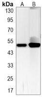
Western blot analysis of p53 (pS6) expression in A549 (A), MCF7 (B) whole cell lysates.
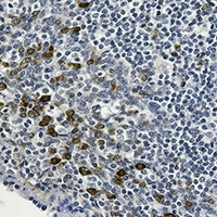
Immunohistochemical analysis of p53 (pS6) staining in human tonsil formalin fixed paraffin embedded tissue section. The section was pre-treated using heat mediated antigen retrieval with sodium citrate buffer (pH 6.126). The section was then incubated with the antibody at room temperature and detected using an HRP conjugated compact polymer system. DAB was used as the chromogen. The section was then counterstained with haematoxylin and mounted with DPX.

关注我们

400-660-9091

电话:025-68037686

地址:江苏生命科技创新园F6幢1层

订购:nanjing03@biogot.com

服务:biorase01@biogot.com

合作:lvyun@biogot.com

支持:may@biogot.com