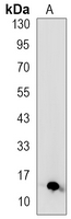
Product Name :S100-A4 Rabbit monoclonal antibody
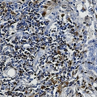
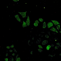
Applications :WB, IHC, IF/ICC, IP
Application_all :WB (1/500 - 1/1000), IHC (1/50 - 1/100), IF/ICC (1/50 - 1/100), IP (1/10 - 1/50)
Background :Despite their relatively small size (8-12 kDa) and uncomplicated architecture, S100 proteins regulate a variety of cellular processes, such as cell growth and motility, cell cycle progression, transcription, and differentiation. To date, 25 members have been identified, including S100A1-S100A18, trichohyalin, filaggrin, repetin, S100P, and S100Z, making it the largest group in the EF-hand, calcium-binding protein family. Interestingly, 14 S100 genes are clustered on human chromosome 1q21, a region of genomic instability. Research studies have demonstrated that significant correlation exists between aberrant S100 protein expression and cancer progression. S100 proteins primarily mediate immune responses in various tissue types but are also involved in neuronal development .Each S100 monomer bears two EF-hand motifs and can bind up to two molecules of calcium (or other divalent cation in some instances). Structural evidence shows that S100 proteins form antiparallel homo- or heterodimers that coordinate binding partner proximity in a calcium-dependent (and sometimes calcium-independent) manner. Although structurally and functionally similar, individual members show restricted tissue distribution, are localized in specific cellular compartments, and display unique protein binding partners, which suggests that each plays a specific role in various signaling pathways. In addition to an intracellular role, some S100 proteins have been shown to act as receptors for extracellular ligands or are secreted and exhibit cytokine-like activities.Research studies have shown that S100A4 is overexpressed in highly metastatic cancers, which makes it useful as a marker of tumor progression and may serve as a prognostic factor in several cancer types . S100A4 exerts its function via direct interaction with a number of proteins including P53, P63, nonmuscle myosin IIA, α6β4 integrin, and liprin b1 . S100A4 is present in the nucleus, cytoplasm and extracellular space. Intracellular and extracellular S100A4 both promote cell migration via interaction with different proteins. Researchers have recently discovered that S100A4 also functions as a neuroprotectant in the peripheral nervous system .
Product :Liquid in 50mM Tris-Glycine (pH 7.4), 0.15M NaCl, 50% Glycerol, 0.01% Sodium azide and 0.05% BSA.
Purification&Purity :The antibody was purified by immunogen affinity chromatography.
Storage&Stability :Store at 4°C short term. Aliquot and store at -20°C long term. Avoid freeze-thaw cycles.
Specificity :Recognizes endogenous levels of S100-A4 protein.
Note :For research use only, not for use in diagnostic procedure.
Alternative Name :CAPL; MTS1; Protein S100-A4; Calvasculin; Metastasin; Placental calcium-binding protein; Protein Mts1; S100 calcium-binding protein A4
Immunogen :A synthetic peptide of human S100A4
Modification :Unmodification