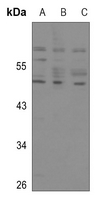
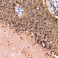
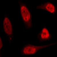
Product Name :p53 polyclonal antibody
Reactivity :Human, Mouse, Rat
Applications :WB, IHC, IF/ICC
Application_all :WB (1/500 - 1/1000), IHC (1/50 - 1/200), IF/ICC (1/50 - 1/200)
Background :The p53 tumor suppressor protein plays a major role in cellular response to DNA damage and other genomic aberrations. Activation of p53 can lead to either cell cycle arrest and DNA repair or apoptosis . p53 is phosphorylated at multiple sites in vivo and by several different protein kinases in vitro. DNA damage induces phosphorylation of p53 at Ser15 and Ser20 and leads to a reduced interaction between p53 and its negative regulator, the oncoprotein MDM2 . MDM2 inhibits p53 accumulation by targeting it for ubiquitination and proteasomal degradation . p53 can be phosphorylated by ATM, ATR, and DNA-PK at Ser15 and Ser37. Phosphorylation impairs the ability of MDM2 to bind p53, promoting both the accumulation and activation of p53 in response to DNA damage . Chk2 and Chk1 can phosphorylate p53 at Ser20, enhancing its tetramerization, stability, and activity .p53 is phosphorylated at Ser392 in vivo and by CAK in vitro . Phosphorylation of p53 at Ser392 is increased in human tumors and has been reported to influence the growth suppressor function, DNA binding, and transcriptional activation of p53 . p53 is phosphorylated at Ser6 and Ser9 by CK1δ and CK1ε both in vitro and in vivo . Phosphorylation of p53 at Ser46 regulates the ability of p53 to induce apoptosis . Acetylation of p53 is mediated by p300 and CBP acetyltransferases. Inhibition of deacetylation suppressing MDM2 from recruiting HDAC1 complex by p19 (ARF) stabilizes p53. Acetylation appears to play a positive role in the accumulation of p53 protein in stress response . Following DNA damage, human p53 becomes acetylated at Lys382 (Lys379 in mouse) in vivo to enhance p53-DNA binding . Deacetylation of p53 occurs through interaction with the SIRT1 protein, a deacetylase that may be involved in cellular aging and the DNA damage response.
Product :Liquid in 0.42% Potassium phosphate, 0.87% Sodium chloride, pH 7.3, 30% glycerol, and 0.01% sodium azide.
Purification&Purity :The antibody was purified by immunogen affinity chromatography.
Storage&Stability :Store at 4°C short term. Aliquot and store at -20°C long term. Avoid freeze-thaw cycles.
Specificity :Recognizes endogenous levels of p53 protein.
Note :For research use only, not for use in diagnostic procedure.
Pathway :AMPK Signaling Pathway,Regulation of Apoptosis,ErbB HER Signaling,Cell Cycle G2 M DNA Damage Signaling Pathway,Mitochondrial Control of Apoptosis,PI3K AKT signaling,Signaling Pathways Activating p38 MAP Kinase,SAPK JNK Signaling Cascades,Warburg Effect.
Alternative Name :P53; Cellular tumor antigen p53; Antigen NY-CO-13; Phosphoprotein p53; Tumor suppressor p53
Immunogen :KLH-conjugated synthetic peptide encompassing a sequence within the N-term region of human p53. The exact sequence is proprietary.
Modification :Unmodification