中国 EN 400-660-9091

联系我们

电话:025-68037686

地址:江苏生命科技创新园F6幢1层

订购:nanjing03@biogot.com

服务:biorase01@biogot.com

合作:lvyun@biogot.com

支持:may@biogot.com

Phospho-TAK1-S412 polyclonal antibody

货号: BS6397
价格 1350
货期 现货(1-2个工作日)
25ul 50ul 100ul 200ul
No. :BS6397
Product Name :Phospho-TAK1-S412 polyclonal antibody
Swiss-Prot :O43318
Host :Rabbit
Reactivity :Human, Mouse, Rat
Applications :WB
Application_all :WB,1:500 - 1:2000
Background :The protein encoded by this gene is a member of the serine/threonine protein kinase family. This kinase mediates the signaling transduction induced by TGF beta and morphogenetic protein (BMP), and controls a variety of cell functions including transcription regulation and apoptosis. In response to IL-1, this protein forms a kinase complex including TRAF6, MAP3K7P1/TAB1 and MAP3K7P2/TAB2; this complex is required for the activation of nuclear factor kappa B. This kinase can also activate MAPK8/JNK, MAP2K4/MKK4, and thus plays a role in the cell response to environmental stresses. Four alternatively spliced transcript variants encoding distinct isoforms have been reported.
Product :1mg/ml in PBS with 0.02% sodium azide, 50% glycerol, pH7.2
Purification&Purity :The antibody was affinity-purified from rabbit antiserum by affinity-chromatography using epitope-specific immunogen and the purity is > 95% (by SDS-PAGE).
Storage&Stability :Store at 4°C short term. Aliquot and store at -20°C long term. Avoid freeze-thaw cycles.
Specificity :Phosphorylated Antibodies
BiowMW :75kDa
Note :For research use only, not for use in diagnostic procedure.
Alternative Name :CSCF;FMD2;MEKK7;TAK1;TGF1a;MAP3K7
Immunogen :A synthetic phosphorylated peptide around S412 of human TAK1(NP_003179.1).
Conjugate :Unconjugated
Modification :Phosphorylated
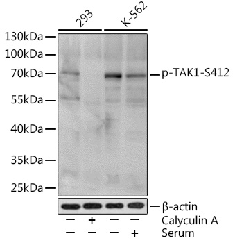
Western blot analysis of extracts of various cell lines, using Phospho-TAK1-S412 Rabbit pAb at 1:1000 dilution.293 cells were treated by Calyculin A at 37℃ for 30 minutes after serum-starvation overnight.K-562 cells were treated by 10% FBS at 37℃ for 30 minutes after serum-starvation overnight.
Secondary antibody: HRP Goat Anti-Rabbit IgG at 1:10000 dilution.
Lysates/proteins: 25ug per lane.
Blocking buffer: 3% BSA.
Detection: ECL Basic Kit .
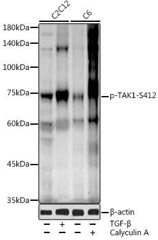
Western blot analysis of extracts of various cell lines, using Phospho-TAK1-S412 antibody at 1:1000 dilution.C2C12 cells were treated by TGF-β at 37℃ for 30 minutes.C6 cells were treated by Calyculin A at 37℃ for 30 minutes after serum-starvation overnight.
Secondary antibody: HRP Goat Anti-Rabbit IgG at 1:10000 dilution.
Lysates/proteins: 25ug per lane.
Blocking buffer: 3% nonfat dry milk in TBST.
Detection: ECL Enhanced Kit .
Exposure time: 120s.

关注我们

400-660-9091

电话:025-68037686

地址:江苏生命科技创新园F6幢1层

订购:nanjing03@biogot.com

服务:biorase01@biogot.com

合作:lvyun@biogot.com

支持:may@biogot.com