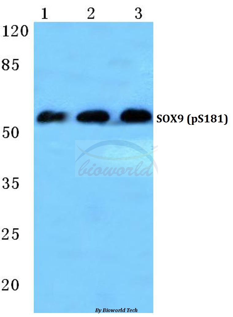
Western blot (WB) analysis of p-SOX9 (S181) polyclonal antibody at 1:500 dilution
Lane1:Hela lysate treated with PMA(100nM,30min)
Lane2:Mouse brain tissue lysate
Lane3:Rat liver tissue lysate
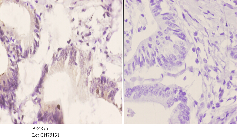
Immunohistochemistry (IHC) analyzes of p-SOX9 (S181) pAb in paraffin-embedded human colon carcinoma tissue at 1:50.showing nucleus staining. Negative control (the right)Using PBS instead of primary antibody, secondary antibody is Goat Anti-Rabbit IgG-biotin followed by avidin-peroxidase.
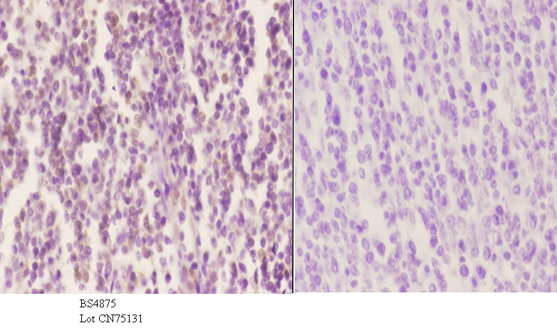
Immunohistochemistry (IHC) analyzes of p-SOX9 (S181) pAb in paraffin-embedded human tonsil carcinoma tissue at 1:50.showing nucleus staining. Negative control (the right)Using PBS instead of primary antibody, secondary antibody is Goat Anti-Rabbit IgG-biotin followed by avidin-peroxidase.