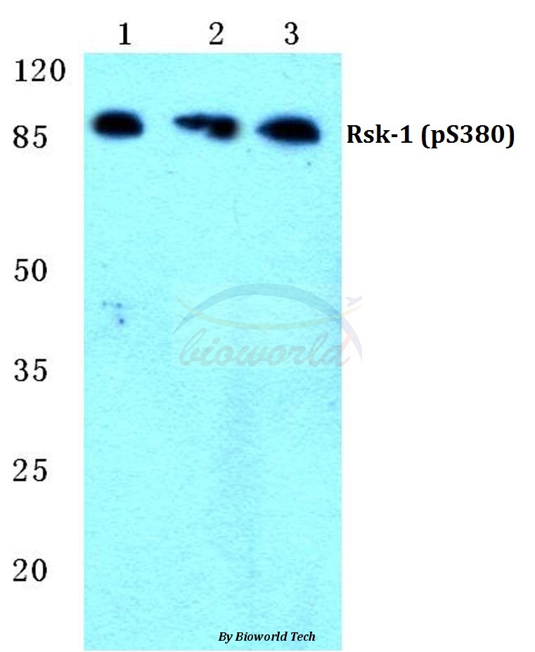
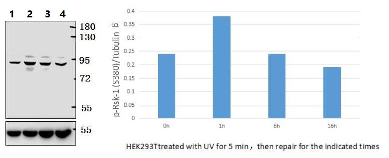
Western blot (WB) analysis of p-Rsk-1 (S380) polyclonal antibody at 1:500 dilution
Lane1:Hela cell lysate treated with PMA(100nM,15mins)
Lane2:NIH-3T3 cell lysate treated with PMA(100nM,15mins)
Lane3:PC12 cell lysate treated with PMA(100nM,15mins)
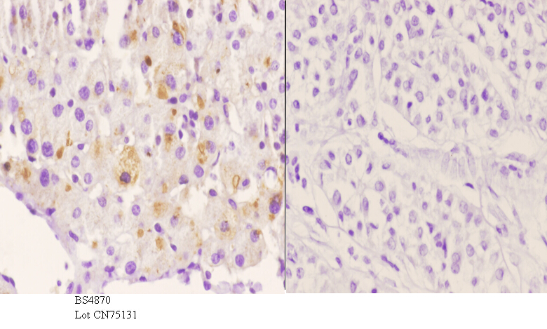
Immunohistochemistry (IHC) analyzes of p-Rsk-1 (S380) pAb in paraffin-embedded human liver carcinoma tissue at 1:50.showing cytoplasmic and nucleus staining. Negative control (the right)Using PBS instead of primary antibody, secondary antibody is Goat Anti-Rabbit IgG-biotin followed by avidin-peroxidase.
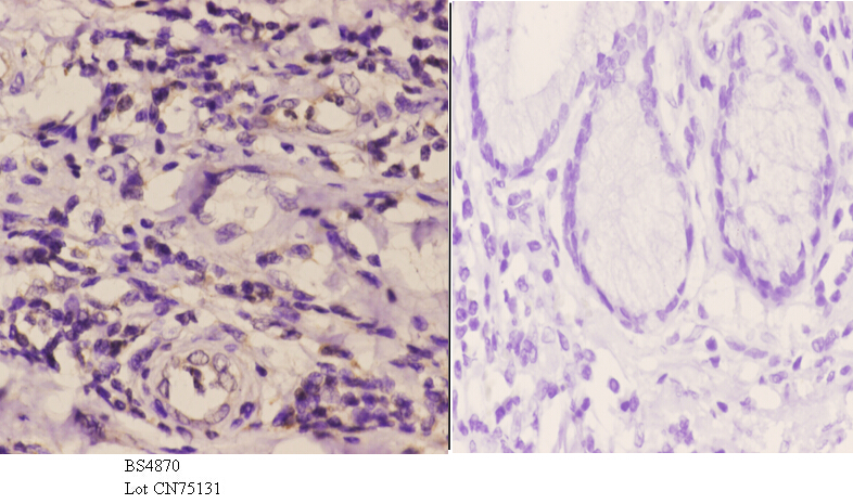
Immunohistochemistry (IHC) analyzes of p-Rsk-1 (S380) pAb in paraffin-embedded human esophageal carcinoma tissue at 1:50.showing cytoplasmic and nucleus staining. Negative control (the right)Using PBS instead of primary antibody, secondary antibody is Goat Anti-Rabbit IgG-biotin followed by avidin-peroxidase.
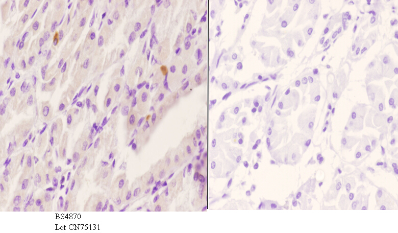
Immunohistochemistry (IHC) analyzes of p-Rsk-1 (S380) pAb in paraffin-embedded human stomach carcinoma tissue at 1:50.showing cytoplasmic and nucleus staining. Negative control (the right)Using PBS instead of primary antibody, secondary antibody is Goat Anti-Rabbit IgG-biotin followed by avidin-peroxidase.