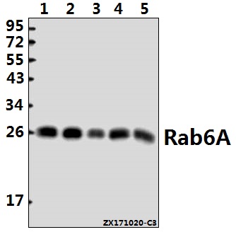
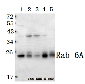
Western blot (WB) analysis of Rab 6A (K144) pAb at 1:500 dilution
Lane1:MCF-7 whole cell lysate(40ug)
Lane2:HepG2 whole cell lysate(40ug)
Lane3:K562 whole cell lysate(40ug)
Lane4:The Testis tissue lysate of Mouse(40ug)
Lane5:The Testis tissue lysate of Rat(40ug)
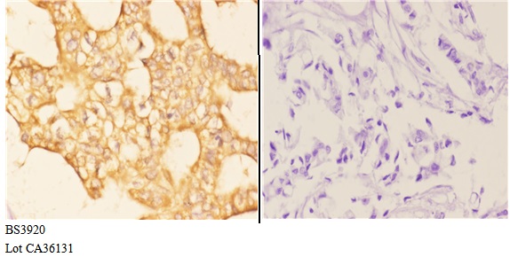
Immunohistochemistry (IHC) analyzes of Rab 6A (K144) pAb in paraffin-embedded human breast carcinoma tissue at 1:50,showing cytoplasmic and membranous staining.Negative control (the right)Using PBS instead of primary antibody, secondary antibody is Goat Anti-Rabbit IgG-biotin followed by avidin-peroxidase.
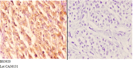
Immunohistochemistry (IHC) analyzes of Rab 6A (K144) pAb in paraffin-embedded human liver carcinoma tissue at 1:50,showing cytoplasmic and membranous staining.Negative control (the right)Using PBS instead of primary antibody, secondary antibody is Goat Anti-Rabbit IgG-biotin followed by avidin-peroxidase.
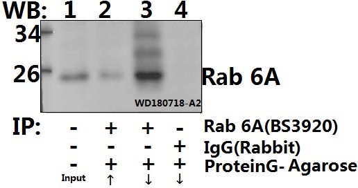
Immunoprecipitation of HEK293T cell lysate using Rab 6A (K144) pAb (Sepharose Bead Conjugate) #BD0048(lane 2 and lane 3) and Nonspecific IgG Control (Sepharose Bead Conjugate)#BD0048 (lane 4 ) .Lane 1 is 30% input. The western blot was probed using Rab 6A (K144).“↑”(supernatant); “↓(deposition)