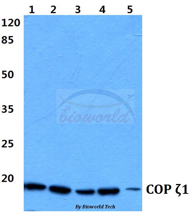
Western blot (WB) analysis of COP ζ1 (I48) polyclonal antibody at 1:500 dilution
Lane1:Hela cell lysate
Lane2:HEK293T cell lysate
Lane3:sp2/0 cell lysate
Lane4:H9C2 cell lysate
Lane5:PC12 cell lysate
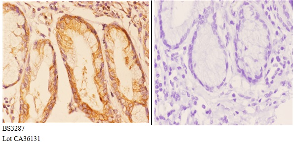
Immunohistochemistry (IHC) analyzes of COP ζ1 (I48) pAb in paraffin-embedded human esophageal carcinoma tissue at 1:50.showing Nucleus speckle and Cytoplasm staining. Negative control (the right)Using PBS instead of primary antibody, secondary antibody is Goat Anti-Rabbit IgG-biotin followed by avidin-peroxidase.
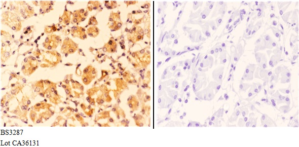
Immunohistochemistry (IHC) analyzes of COP ζ1 (I48) pAb in paraffin-embedded human stomach carcinoma tissue at 1:50.showing Nucleus speckle and Cytoplasm staining. Negative control (the right)Using PBS instead of primary antibody, secondary antibody is Goat Anti-Rabbit IgG-biotin followed by avidin-peroxidase.