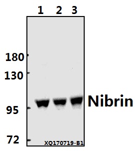
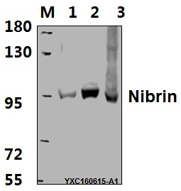
Western blot (WB) analysis of Nibrin (P461) pAb at 1:1000 dilution
Lane1:HEK293T whole cell lysate(40ug)
Lane2:HepG2 whole cell lysate(40ug)
Lane3:LO2 whole cell lysate(40ug)
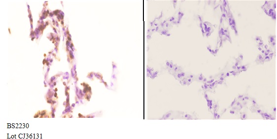
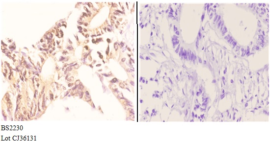
Immunohistochemistry (IHC) analyzes of Nibrin (P461) pAb in paraffin-embedded lung carcinoma tissue at 1:50 showing Nucleus staining. Negative control (the right)Using PBS instead of primary antibody, secondary antibody is Goat Anti-Rabbit IgG-biotin followed by avidin-peroxidase.
Immunohistochemistry (IHC) analyzes of Nibrin (P461) pAb in paraffin-embedded lung carcinoma tissue at 1:50 showing Nucleus staining. Negative control (the right)Using PBS instead of primary antibody, secondary antibody is Goat Anti-Rabbit IgG-biotin followed by avidin-peroxidase.