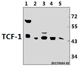
Western blot (WB) analysis of TCF-1 (F26) pAb at 1:500 dilution
Lane1:K562 whole cell lysate(40ug)
Lane2:A549 whole cell lysate(40ug)
Lane3:U-87MG whole cell lysate(40ug)
Lane4:C6 whole cell lysate(40ug)
Lane5:MEF whole cell lysate(40ug)
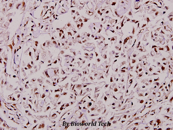
Immunohistochemistry (IHC) analyzes of TCF-1 (F26) pAb in paraffin-embedded human breast carcinoma tissue at 1:100.
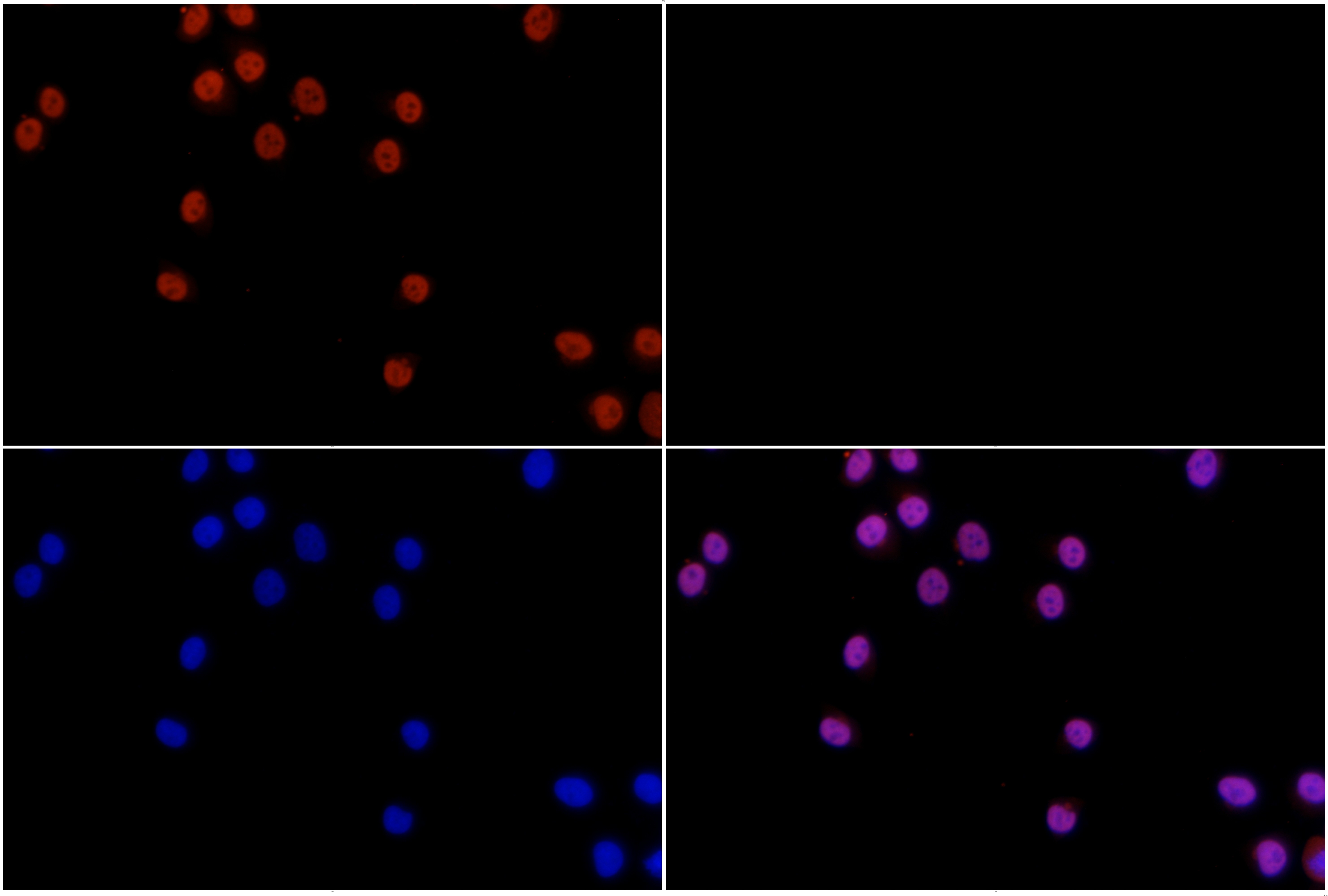
Immunofluorescence analysis of L02 cells using TCF-1 (F26) pAb at dilution of 1:200 (40x lens).
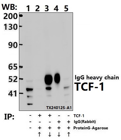
Immunoprecipitation of HCT116 cell lysates using TCF-1 pAb (Sepharose Bead Conjugate)#BD0048 (lane 2 and lane 3) and Nonspecific IgG Control (Sepharose Bead Conjugate)#BD0048 (lane 4 and lane 5) .Lane 1 is 30% input. The western blot was probed using TCF-1 pAb.