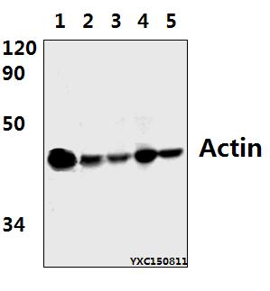
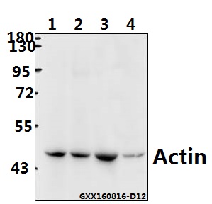
Western blot (WB) analysis of Actin (E361) polyclonal antibody at 1:500 dillution
Lane1:Hela whole cell lysate(40μg)
Lane2:HEK293T whole cell lysate(40μg)
Lane3:NIH-3T3 whole cell lysate(40μg)
Lane4:RAW264.7 whole cell lysate(40μg)
Lane5:PC12 whole cell lysate(40μg)
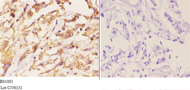
Immunohistochemistry (IHC) analyzes of Actin (E361) pAb in paraffin-embedded human breast carcinoma tissue at 1:50.showing cytoplasmic and cytoskeleton staining. Negative control (the right)Using PBS instead of primary antibody, secondary antibody is Goat Anti-Rabbit IgG-biotin followed by avidin-peroxidase.
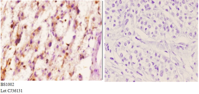
Immunohistochemistry (IHC) analyzes of Actin (E361) pAb in paraffin-embedded human liver carcinoma tissue at 1:50.showing cytoplasmic and cytoskeleton staining. Negative control (the right)Using PBS instead of primary antibody, secondary antibody is Goat Anti-Rabbit IgG-biotin followed by avidin-peroxidase.