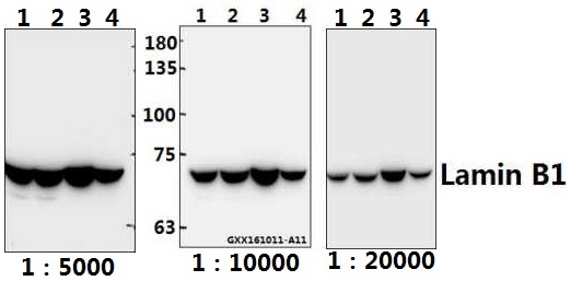
Western blot (WB) analysis of Lamin B1 polyclonal antibody at 1:20000 dilution
Lane1:A549 whole cell lysate(40ug)
Lane2:DLD whole cell lysate(40ug)
Lane3:The Spleen tissue lysate of Mouse(40ug)
Lane4:The Lung tissue lysate of Rat(40ug)
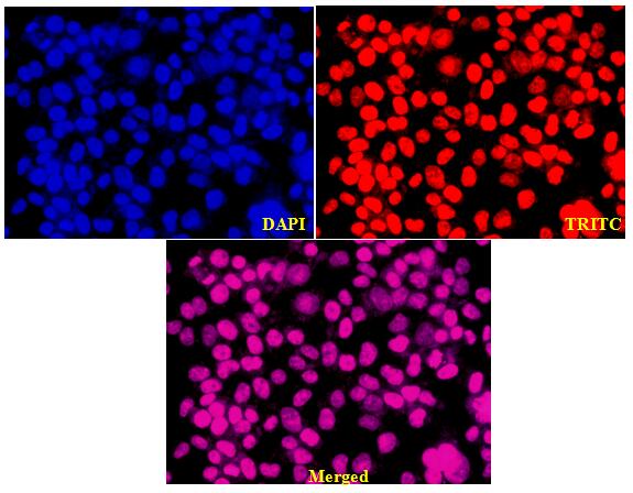
Immunofluores image of AP6001 stained HEK293T cells. The cells were 4% paraformaldehyde fixed (20 min) and then incubated in 10% normal goat serum for 1h to permeabilise the cells and block non-specific protein-protein interactions. The cells were then incubated with the antibody Lamin B1 pAb #AP6001(1:200) at 5µg/ml overnight at +4°C. The secondary antibody (Red) was Goat Anti-Rabbit IgG (H+L) Rhodamine (TRITC) #BS10250 used at a 1/2000 dilution for 1h. DAPI #BD5014 was used to stain the cell nuclei (blue).
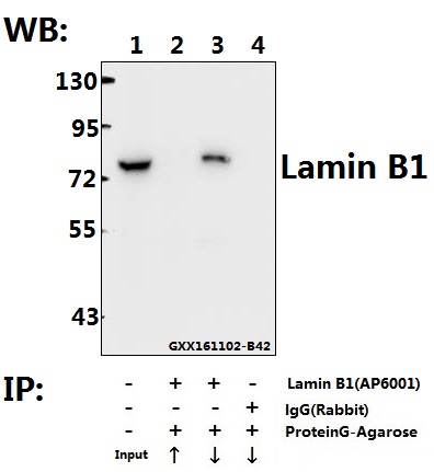
Immunoprecipitation of the Lung tissue lysate of Mouse using Lamin B1 pAb (Sepharose Bead Conjugate) #BD0047(lane 2 and lane 3) and Nonspecific IgG Control (Sepharose Bead Conjugate)#BD0047 (lane 4) .Lane 1 is 30% input. The western blot was probed using Lamin B1 pAb #AP6001.“↑”(supernatant); “↓”(deposition)