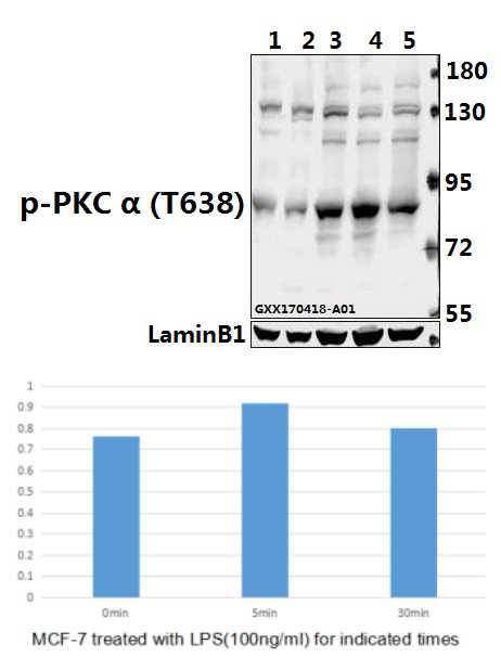
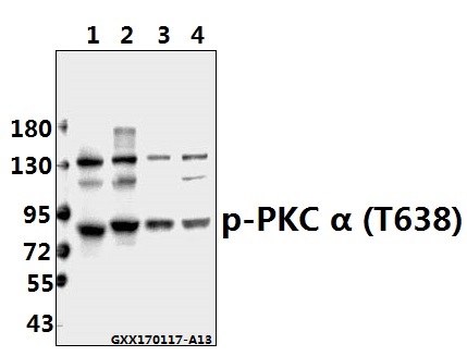
Western blot (WB) analysis of PKC α (phospho-T638) polyclonal antibody at 1:500 dilution
Lane1:PC12 whole cell lysate(40ug)
Lane2:CT26 whole cell lysate(40ug)
Lane3:MCF-7 treated with LPS(100ng/ml) for 30 minutes whole cell lysate
Lane4:MCF-7 treated with LPS(100ng/ml) for 5 minutes whole cell lysate
Lane5:MCF-7 whole cell lysate
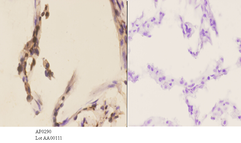
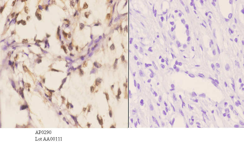
Immunohistochemistry (IHC) analyzes of p-PKC α (T638) pAb in paraffin-embedded human lung carcinoma tissue at 1:50.showing cytoplasmic and nucleus staining. Negative control (the right)Using PBS instead of primary antibody, secondary antibody is Goat Anti-Rabbit IgG-biotin followed by avidin-peroxidase.
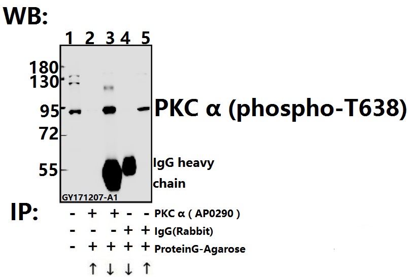
Immunoprecipitation of L02 cell lysate using PKC α (phospho-T638) pAb (Sepharose Bead Conjugate) #BD0048(lane 2 and lane 3) and Nonspecific IgG Control (Sepharose Bead Conjugate)#BD0048 (lane 4) .Lane 1 is 30% input. The western blot was probed using SHP-2 (E574) pAb.“↑”(supernatant); “↓”(deposition)