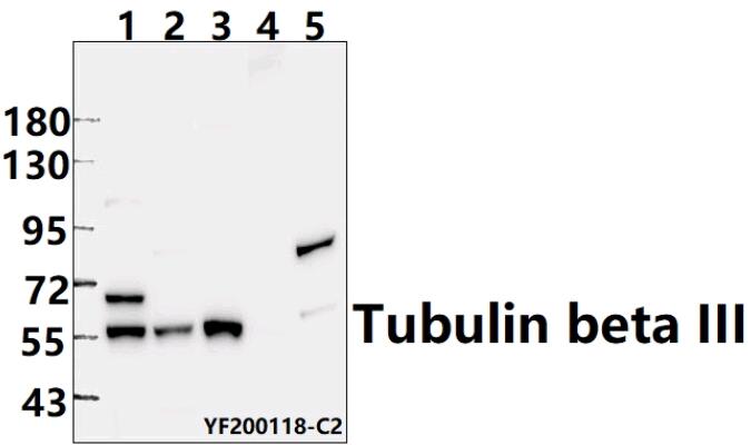
Western blot (WB) analysis of Tubulin beta III pAb at 1:500 dilution
Lane1:A549 whole cell lysate(40ug)
Lane2:PC12 whole cell lysate(40ug)
Lane3:MEF whole cell lysate(40ug)
Lane4:The kidney tissue lysate of Mouse(40ug)
Lane5:The lung tissue lysate of Rat(40ug)
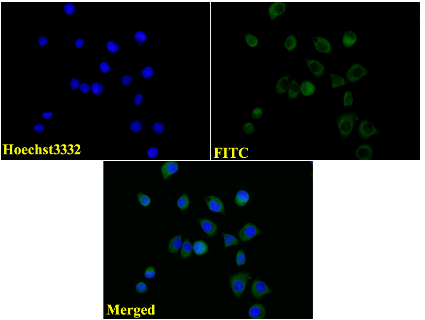
IF image of AP0013 stained A549 cells.The cells were 4% paraformaldehyde fixed (20 min) and then incubated in 10% normal goat serum for 1h to permeabilise the cells and block non-specific protein-protein interactions. The cells were then incubated with the antibody Tubulin beta III #AP0013(1:100) at 10µg/ml overnight at +4°C. The secondary antibody (Green) was Goat Anti-Rabbit IgG (H+L) FITC #BS10950 used at a 1/1000 dilution for 1h. Hoechst33342 #BD5011 was used to stain the cell nuclei (blue).